MRI Scan
Magnetic resonance imaging (MRI) is a noninvasive radiological scan to examine your organs, tissues, and skeletal system. It produces high-resolution images of the inside of the body that help diagnose a variety of problems. The resulting 3D images can be viewed from many different angles.
The content has been reviewed for quality and accuracy to the best of our knowledge by Qunomedical and its Medical Board of Experts.
How Does an MRT Work?
MRI does not involve X-rays, the use of ionizing radiation or radioactive tracers, which distinguishes it from CT, CAT, and PET scans.
An MRI scan can be used to examine almost any part of the body, including:
brain and spinal cord
heart and blood vessels
internal organs
bones and joints
breasts
The results of an MRI scan can be used to help diagnose conditions, plan treatments and assess how effective previous treatment has been.
What Happens During an MRI Scan?
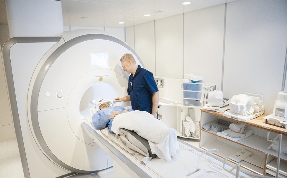
MRI scanners are large tubes that contain powerful magnets. You lie inside the tube on a flat bed that's moved into the scanner.
The scan lasts 15 to 90 minutes, depending on the size of the area being scanned and how many images are taken. Depending on the part of your body being scanned, you'll be moved into the scanner either head first or feet first. It's very important to keep as still as possible during your MRI scan, so that accurate images can be obtained.
The majority of the human body is made of water. Water or H2O, is made up of hydrogen protons and the MRI machines exploit the abundance of these hydrogen protons in your body. Inside the MRI machine, the magnetic field temporarily realigns hydrogen atoms in your body. Radio waves produce a varying magnetic field, which results in these atoms changing their alignment and in the process produce very faint signals, which are used to create MRI images
The scanner is operated by a radiographer, who is trained in carrying out imaging investigations. They control the scanner using a computer, which is in a different room, to keep it away from the magnetic field generated by the scanner. You can talk to the radiographer through an intercom during the scan and they'll be able to see you on a television monitor throughout the scan. The scanner will make very loud tapping noises, but don’t worry, this is the electric current in the scanner coils being turned on and off. Headphones are usually provided to mask the sound.
What Are the Different Applications of MRI?
MRI of the Brain and Spinal Cord
MRI provides excellent imaging of the brain, therefore making it the tool of choice for investigating neurological cancers. Due to the high level visualization of grey and white matter in the brain, MRI is used to diagnose conditions of the Central Nervous System (CNS), including dementia, Multiple Sclerosis, Alzheimer’s disease and epilepsy.
MRI of the Heart and Blood Vessels
MRI is used in cardiology diagnostics alongside other imaging techniques to assess conditions such as vascular diseases and congenital heart disease.
MRI of Internal Organs
MRI is also used to diagnose conditions of the internal organs such as identifying small bowel tumors inflammatory bowel disease, tumors in the liver, and any abnormalities in the kidney, pancreas. or conditions of the arteries and blood vessels, such as stenosis or aneurysms. Contrast agents may be used in combination to provide accurate images.
MRI of Bones and Joints
In orthopedics, MRI is used to examine bones, joints and soft tissue to detect the presence of abnormalities such as tumors, inflammatory disease, and spinal cord disc degeneration.
Other Applications
MRI is also used to detect breast cancer and other abnormalities in the breast. It will usually be requested after a positive biopsy sample has been taken, so the physician can get more information about the extent of the disease.For women at high risk for breast cancer, a screening MRI is recommended along with a yearly mammogram. Reproductive organs and other pelvic organs can also be viewed.
Is MRI Safe?
MRI is widely used in hospitals and clinics for medical diagnosis, staging of disease and follow-up without exposing the body to radiation, and may be considered as a better choice than a CT scan.
In general, there is no known risk with MRI. It is even safe for pregnant women, as X-Rays are not used. People with medical implants or other non-removable metal inside the body however, must consult their physician beforehand, as foreign material can significantly distort the images taken from the scanner.
Finally, if you do not like being in enclosed spaces, you may need to take medicine to help you relax while in the scanner. Although you will be alone in the exam room, you can talk to the MR technologist, who can see and hear what’s going on.
MRI Scan Costs
Below, we've laid out MRI scan starting costs in a number of different locations. Bear in mind though that these are not fixed or guaranteed and may vary based on a number of different factors.
| Country | Price (EUR €) |
|---|---|
| Hungary | €197 |
| Poland | €198 |
| Lithuania | €330 |
| Austria | €500 |
| Turkey | €536 |
| Germany | €800 |

Patient manager
Frieda
Your personal Patient Manager
Let's talk
Still unsure? Feeling overwhelmed? Talking to a real person can give you the guidance and reassurance needed. You don’t have to do it alone. Let’s find the right doctor together.